
Serratus posterior superior muscle
This article includes a list of references, related reading, or external links, but its sources remain unclear because it lacks inline citations. (May 2015) |
| Serratus posterior superior muscle | |
|---|---|
 Thin film-like object, at center, is serratus posterior superior muscle. | |
| Details | |
| Origin | Nuchal ligament (or ligamentum nuchae) and the spinous processes of the vertebrae C7 through T3 |
| Insertion | The upper borders of the 2nd through 5th ribs |
| Artery | Intercostal arteries |
| Nerve | 2nd through 5th intercostal nerves |
| Actions | Elevates ribs 2-5 [1] |
| Identifiers | |
| Latin | musculus serratus posterior superior |
| TA98 | A04.3.01.011 |
| TA2 | 2236 |
| FMA | 13401 |
| Anatomical terms of muscle | |
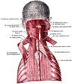
The serratus posterior superior muscle is a thin, quadrilateral muscle. It is situated at the upper back part of the thorax, deep to the rhomboid muscles.
Structure[edit]
The serratus posterior superior muscle arises by an aponeurosis from the lower part of the nuchal ligament, from the spinous processes of C7, T1, T2, and sometimes T3, and from the supraspinal ligament.[2] It is inserted, by four fleshy digitations into the upper borders of the second, third, fourth, and fifth ribs past the angle of the rib.[2]
Function[edit]
The serratus posterior superior muscle elevates the second to fifth ribs.[citation needed] This aids deep respiration.[citation needed]
Additional images[edit]
-
 Position of serratus posterior superior muscle (shown in red).
 -
 Serratus posterior superior muscles are labeled at center left and center right.


See also[edit]
References[edit]
- ^ According to Moore et al (Moore Clinically Oriented Anatomy 7th Edition Chapter 1: Thorax, page 86) and Vilensky et al (Clin Anat. 2001 Jul;14(4):237-41. Serratus posterior muscles: anatomy, clinical relevance, and function. Vilensky JA, Baltes M, Weikel L, Fortin JD, Fourie LJ : The serratus posterior superior and inferior muscles are generally considered clinically insignificant muscles that, based on attachments, probably function in respiration. However, there is no evidence supporting a respiratory role for these muscles. In fact, some electromyographic data refute a respiratory function for these muscles. We suggest that the serratus posterior muscles function primarily in proprioception. Further, these muscles, especially the superior, have been implicated in myofascial pain syndromes and therefore may have greater clinical relevance than commonly attributed to them.
- ^ a b Jolley, C. J.; Moxham, J. (January 1, 2006), "RESPIRATORY MUSCLES, CHEST WALL, DIAPHRAGM, AND OTHER", in Laurent, Geoffrey J.; Shapiro, Steven D. (eds.), Encyclopedia of Respiratory Medicine, Oxford: Academic Press, pp. 632–643, ISBN 978-0-12-370879-3, retrieved January 17, 2021
![]() This article incorporates text in the public domain from page 404 of the 20th edition of Gray's Anatomy (1918)
This article incorporates text in the public domain from page 404 of the 20th edition of Gray's Anatomy (1918)
- Clinically Oriented Anatomy, 4th ed. Keith L. Moore and Arthur F. Dalley.
- Board Review Series: Gross Anatomy, 4th ed. Kyung Won Chung.
External links[edit]
- Anatomy figure: 01:05-02 at Human Anatomy Online, SUNY Downstate Medical Center - "Intermediate layer of the extrinsic muscles of the back, deep muscles."